HEARING CARE
Understanding Ear Anatomy: Parts and Their Functions
By Team Hearzap | Oct. 3, 2025
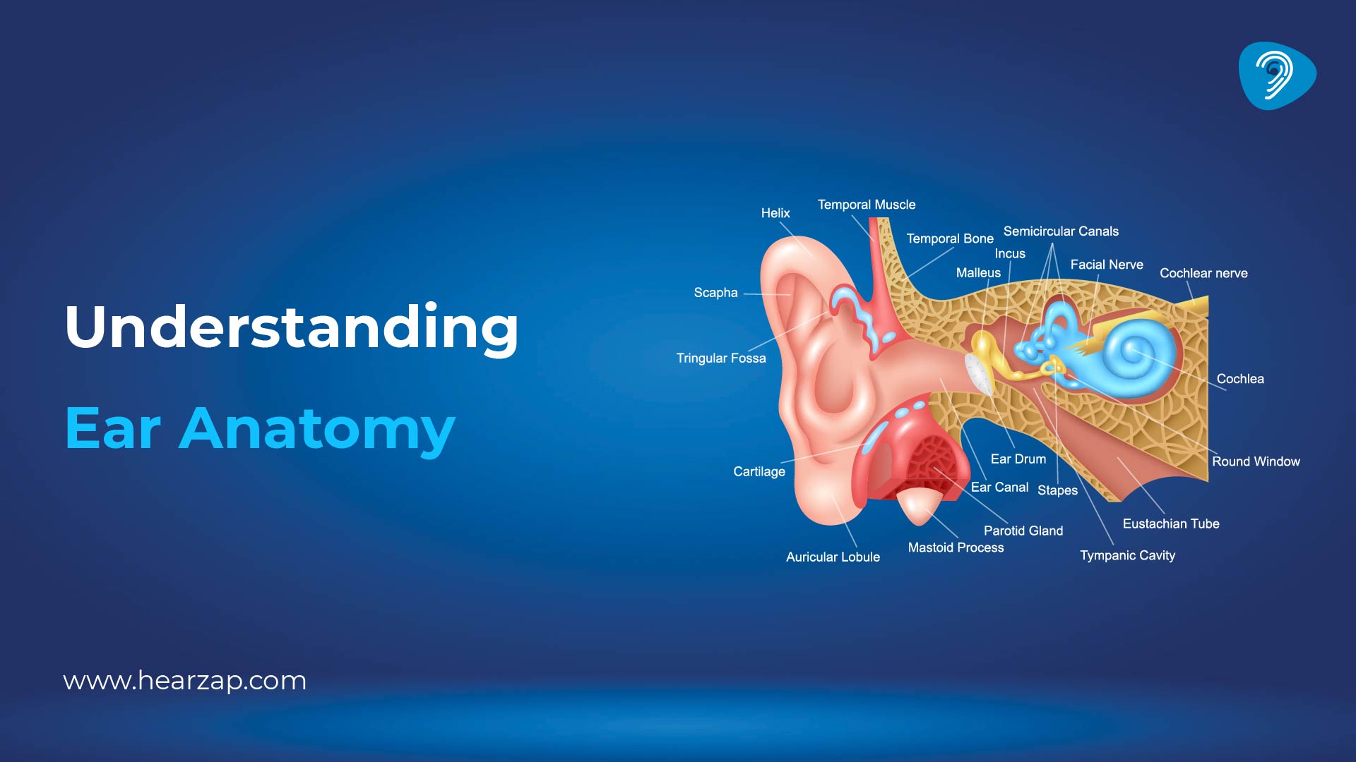
Our ears help us communicate, keep our balance and protect delicate tissues from the world outside. A clear grasp of ear anatomy gives you a simple map of what sits in the outer, middle and inner sections, and how those pieces work together in everyday life. This article explores the three main parts of the ear, breaking down their roles in capturing sound, transmitting it, and translating it into signals the brain understands.
Overview: Parts of the Ear
Doctors and audiologists talk about three parts of the ear that connect in sequence. The outer section collects sound and shields the canal. The middle section carries vibration and manages pressure. The inner section converts vibration into electrical messages and senses head movement. Inside those regions are smaller ear parts with specific jobs. When the chain is smooth, everyday ear functions feel effortless.
Anatomy of the Outer (External) Ear
Function of the External Ear
The outer ear is a collector and a guard. The curved pinna acts like a funnel and nudges sound into the canal, which slightly boosts some speech frequencies. You can test the idea by cupping a hand behind your ear while someone speaks softly. The shape gathers more energy, so the voice seems clearer. The canal’s skin makes wax that traps dust and carries it slowly outward. Tiny hairs near the entrance form a barrier. Together, these features guide sound in and keep unwanted particles out.
Structure & Components
The outer ear is made up of three main parts - the pinna, the ear canal, and the eardrum.
1. Pinna (Auricle):
The pinna, also known as the auricle, is the visible part of the ear made of flexible cartilage covered by skin. Its ridges and curves help capture sound waves and shape them based on direction, whether they come from the front, back, or above. This helps the brain figure out where a sound is coming from.
2. Ear Canal:
Next is the ear canal, a slightly curved passage that leads sound towards the eardrum. The outer third of the canal is supported by cartilage, while the inner section is anchored to bone. Together, they protect the eardrum and guide sound efficiently.
3. Eardrum (Tympanic Membrane):
At the end of the canal lies the eardrum - a thin, pearl-grey membrane that separates the outer ear from the middle ear. It vibrates in response to incoming sound waves, converting them into mechanical energy that continues through the hearing system.
Acoustic Note:
The ear canal acts like a small resonator, roughly 2.5 centimetres long. Given that the speed of sound in air is about 340 metres per second, this length naturally amplifies sounds near 3.4 kilohertz - a frequency range that matches key tones used in many Indian languages.
Anatomy of the Middle Ear
Function of the Middle Ear
The middle ear acts as a bridge between the outer and inner ear. Its main role is to carry the vibrations from the eardrum into the fluid-filled inner ear with minimal energy loss. It achieves this through a system of three tiny bones and a pressure-balancing tube.
The three bones work together as a lever system, transferring sound from the large surface area of the eardrum to the much smaller oval window. This concentration of force makes sound transmission more efficient. Two small muscles also help protect the ear during loud sounds by tightening the chain of bones for a brief moment.
The Eustachian tube, or pressure tube, connects the middle ear to the back of the nose. It opens briefly when you swallow, yawn, or chew, ensuring that air pressure remains equal on both sides of the eardrum.
Structure & Components
The front wall of the middle ear is formed by the eardrum, which separates it from the outer ear. Behind the eardrum lies a chain of three tiny bones known as the ossicles:
- Malleus (Hammer): Attached to the inner surface of the eardrum, it receives the initial vibration.
- Incus (Anvil): Connects the malleus to the stapes.
- Stapes (Stirrup): The smallest bone in the human body, it pushes on the oval window, transmitting the vibrations into the inner ear’s fluid.
Together, these bones convert air vibrations into fluid vibrations that the inner ear can interpret.
Two small muscles provide control and protection:
- The tensor tympani tightens the eardrum by pulling on the malleus.
- The stapedius limits the movement of the stapes during sudden loud sounds to prevent damage.
The Eustachian tube slopes downward to connect the middle ear with the back of the nose. It stays closed most of the time but opens during swallowing or yawning to balance pressure. Because children have shorter and more horizontal tubes, they are more likely to experience fluid buildup behind the eardrum. Adults may also feel ear pressure or fullness during colds or sinus infections for the same reason.
Anatomy of the Inner Ear
Function of the Inner Ear
The inner ear has two main roles: hearing and balance. It converts sound vibrations into electrical signals that the brain recognises as sound, and it senses head movement to help maintain balance.
The cochlea, a spiral, fluid-filled structure, handles hearing. When the stapes pushes on the oval window, it creates waves in the cochlear fluid. A flexible strip called the basilar membrane vibrates at different spots for high and low sounds. Hair cells on this membrane detect the motion and send signals through the auditory nerve to the brain.
Structure & Components
The cochlea contains three spiral fluid chambers. Sitting on the basilar membrane, the organ of Corti holds inner and outer hair cells that bend with fluid movement. Damage to these cells from loud noise or certain medicines can lead to hearing loss.
Beside the cochlea is the balance system, which includes three semicircular canals and two sacs called the utricle and saccule. The canals detect rotation, while the sacs sense tilt and straight-line motion. Tiny crystals inside these sacs move with your head, bending hair cells and sending balance signals through the vestibular nerve to the brain.
How Ear Anatomy Relates to Ear Health
Knowing the anatomy helps you choose safe habits and know when to seek help. Think of three rooms in one house, where a leak in one can affect the others.
Everyday Care For The Outer Ear
- Avoid digging deep with cotton buds, keys or hairpins. The canal skin is thin, and the eardrum sits closer than you think. Pushing wax inwards is a common reason for blockage.
- After a swim or shampoo, tilt your head to let water drain.
- If you notice itch, pain or discharge, resist scratching and read about ear infections. A clinician can examine the canal with light and advise the next steps.
Looking After The Middle Ear
- During flights or hill travel, swallow, sip water or yawn during take-off and landing to open the Eustachian tube.
- Treat colds and allergies sensibly. A blocked nose often means a blocked tube, which can lead to fluid behind the eardrum and a heavy feeling.
- If sound feels muffled after a cold, give it a few days, then seek a review if it lingers.
Caring For The Inner Ear And Balance
- Use safe listening habits. Keep personal audio at a comfortable level and take listening breaks.
- Report sudden spinning, one-sided ringing or a hearing drop quickly.
When To Consider A Hearing Check
A hearing test shows how sound travels through the outer and middle sections and how clearly the inner section and the nerve send signals. People book appointments for many reasons, for example, difficulty following speech in noise, a high television volume or ringing in silence. Tests also help monitor ear health before and after noisy work. If results suggest a concern, the report will outline options involving hearing loss or ear infections.
Linking Symptoms To Structure
- Pain at the entrance or an itch that worsens after swimming often points to canal skin irritation, an outer ear issue.
- A sense of fullness with recent cold symptoms may suggest Eustachian tube blockage in the middle section.
- Spinning with nausea tends to involve the inner ear balance organs.
These patterns are not diagnoses. They simply show how the map of ear anatomy connects to daily experiences.
Conclusion
A simple understanding of ear anatomy turns vague worries into practical steps. The outer section collects and protects. The middle section transmits vibration and balances pressure. The inner section converts motion into nerve signals and senses movement for balance. When these rooms are clear in your mind, decisions about cleaning habits, travel, safe listening and when to book a hearing test become easier.
FAQs
What is the basic anatomy of the ear?
The ear has three regions. The outer section includes the pinna, ear canal and eardrum. The middle section holds the ossicles and the Eustachian tube. The inner section contains the cochlea for hearing and the vestibular organs for balance. These are the standard parts of the ear shown in clinic posters.
What are the main parts of the ear?
Professionals group them as outer, middle and inner sections of the system. Lists of ear parts usually name the pinna, ear canal, eardrum, malleus, incus, stapes, Eustachian tube, cochlea, semicircular canals, vestibule and auditory nerve. These names appear across guides to external ear anatomy, middle ear anatomy and inner ear anatomy.
How does the inner ear anatomy affect balance and hearing?
Fluid waves in the cochlea bend hair cells, which release signals into the auditory nerve. The semicircular canals sense rotation, and the utricle and saccule sense tilt and straight-line acceleration. That dual role explains why inner ear anatomy appears in both hearing and balance checks.
Can outer ear anatomy problems cause hearing loss?
Yes. If the canal is blocked by wax, swelling or debris, less sound reaches the eardrum and hearing feels dull. Infections of canal skin can also cause pain and a temporary drop in hearing. Clearing the cause often restores clarity, which shows how external ear anatomy links to sound quality and comfort.
How do I keep my ear health good, especially for internal ear parts?
Use safe listening levels, take breaks from loud sound, stay active to keep balance pathways fresh and protect the head during sport or travel. Book a hearing test if you notice changes, ringing or a blocked feeling that does not settle. Simple hearing care tips, such as avoiding deep cleaning with objects and drying the ears gently after a bath, support the whole system.
Related Blogs
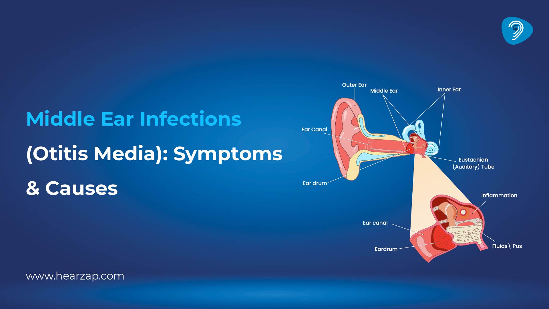
Middle Ear Infections (Otitis Media): Symptoms & Causes
Hyperacusis: Causes of Hearing Sensitivity and Treatment Options
10 Hearing Care Tips & Maintenance
Contact us
We are here for all your hearing needs, from hearing tests to hearing aids. Fill out the form below, and we will give you a call soon.
Recent Blogs
By None | March 6, 2026
By Team Hearzap | March 4, 2026
By Team Hearzap | March 2, 2026
By None | Feb. 25, 2026
By None | Feb. 20, 2026